 When an owner thinks of a mass, regardless of the location on the body, cancer should always come to mind. Yet, not all masses that grow inside or on surface of the body are actually cancerous.
I’m well aware of such varying results in my patients, but the reality of being faced with my own dog potentially having cancer besides that which has been cut out his abdomen twice in the past two years is something I just had to face.
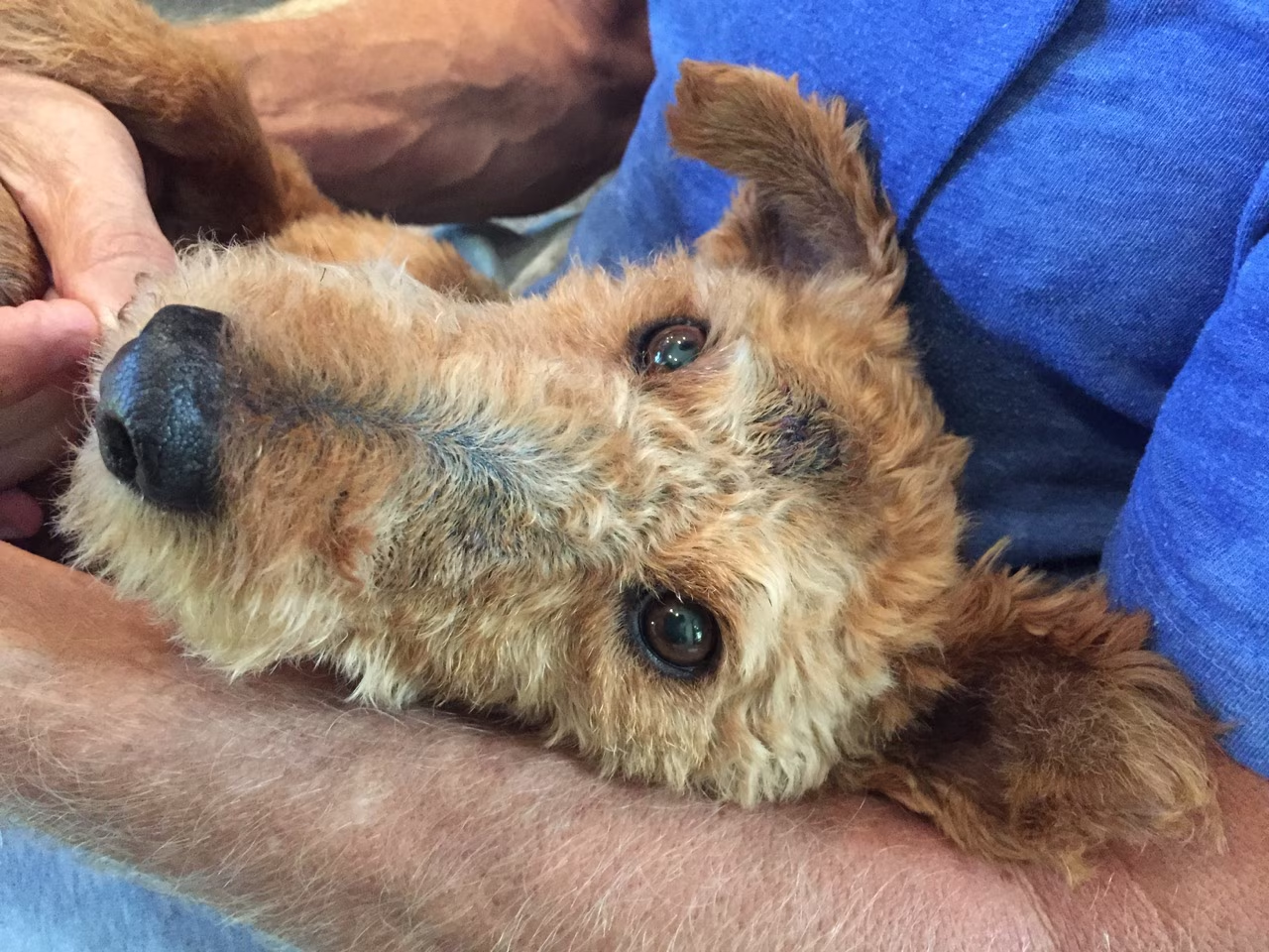
Now that you’ve read about Cardiff’s skin mass removals (see What We Do When There Are Tumors On the Inside and On the Outside), you’re likely wondering about his biopsy results. Well, it’s time for the big reveal. Yes, Cardiff has now been diagnosed with more than one type of cancer. Fortunately, the diagnoses show just one more malignant cancer besides his lymphoma, along with many spots of benign cancer.
Before I reveal Cardiff’s results, let’s cover the differences between benign and malignant cancers. First, what exactly is cancer? Cancer occurs when a cell’s genetic material (DNA) becomes irreversibly damaged, which initiates rapid division that does not turn itself off. When enough cancerous cells have multiplied a mass forms. Neoplasia or neoplasm are other terms used for cancer.
Benign cancer is typically less concerning as it has less of a tendency to spread (metastasize), but can still be locally invasive. Malignant cancer is more concerning, as it has a higher likelihood of metastasizing to other regions of the body through the lymphatic system and blood vessels and can cause considerable damage at its site of origin.
Yet, some masses aren’t cancer at all. Normal cells can divide excessively and form masses (still an abnormal process). Alternatively, fluid filled cysts, inflammatory reactions (like the body’s attempt to wall off a splinter), or others conditions can cause mass-like lesions. That’s why we don’t want to jump to the conclusion that a pet has cancer until the mass has been evaluated by a veterinarian, who has confirmed such suspicions with either a fine needle aspirate (FNA) for cytology or a biopsy.
When surgically removing a benign or non-cancerous mass, there’s less of a need to have wider margins of normal tissue removed in addition to the site of concern. With malignant tumors, it’s crucial that more normal tissue is cut out to ensure the cancer has not invaded deeper into the site of origin on a microscopic level. Two to three centimeters in all directions is an ideal margin for masses suspected or confirmed to be malignant. Surgically removing (excising) a mass with clean margins is often curative.
When an owner thinks of a mass, regardless of the location on the body, cancer should always come to mind. Yet, not all masses that grow inside or on surface of the body are actually cancerous.
I’m well aware of such varying results in my patients, but the reality of being faced with my own dog potentially having cancer besides that which has been cut out his abdomen twice in the past two years is something I just had to face.
Now that you’ve read about Cardiff’s skin mass removals (see What We Do When There Are Tumors On the Inside and On the Outside), you’re likely wondering about his biopsy results. Well, it’s time for the big reveal. Yes, Cardiff has now been diagnosed with more than one type of cancer. Fortunately, the diagnoses show just one more malignant cancer besides his lymphoma, along with many spots of benign cancer.
Before I reveal Cardiff’s results, let’s cover the differences between benign and malignant cancers. First, what exactly is cancer? Cancer occurs when a cell’s genetic material (DNA) becomes irreversibly damaged, which initiates rapid division that does not turn itself off. When enough cancerous cells have multiplied a mass forms. Neoplasia or neoplasm are other terms used for cancer.
Benign cancer is typically less concerning as it has less of a tendency to spread (metastasize), but can still be locally invasive. Malignant cancer is more concerning, as it has a higher likelihood of metastasizing to other regions of the body through the lymphatic system and blood vessels and can cause considerable damage at its site of origin.
Yet, some masses aren’t cancer at all. Normal cells can divide excessively and form masses (still an abnormal process). Alternatively, fluid filled cysts, inflammatory reactions (like the body’s attempt to wall off a splinter), or others conditions can cause mass-like lesions. That’s why we don’t want to jump to the conclusion that a pet has cancer until the mass has been evaluated by a veterinarian, who has confirmed such suspicions with either a fine needle aspirate (FNA) for cytology or a biopsy.
When surgically removing a benign or non-cancerous mass, there’s less of a need to have wider margins of normal tissue removed in addition to the site of concern. With malignant tumors, it’s crucial that more normal tissue is cut out to ensure the cancer has not invaded deeper into the site of origin on a microscopic level. Two to three centimeters in all directions is an ideal margin for masses suspected or confirmed to be malignant. Surgically removing (excising) a mass with clean margins is often curative.
What Were the Results From Cardiff’s Biopsies?
My hope was that the majority of his skin masses were benign tumors or even potentially non-cancerous lesions. Considering we veterinarians are often blessed (or is it cursed?) by having personal pets with unusual medical histories, the trend continues for Cardiff. It turns out that Cardiff had a combination of six types of cancerous and noncancerous masses. Six of nine were cancerous, with four being benign and two being malignant. Here's the breakdown of the biopsy results with explanations as per Idexx Laboratories, which is the diagnostic testing service I use for my patients: 1. Sebaceous adenoma Cardiff had three sebaceous adenomas, which were on the right side of his head, the right shoulder, and on the outside of his right hind limb. “Sebaceous gland adenomas are the most common epithelial skin tumors in the dog and are also seen occasionally in cats. They are most common in older animals and may involve the skin anywhere on the body. They are usually very well circumscribed and have a low potential for local recurrence after complete surgical excision.” So, the sebaceous adenoma is a benign skin tumor. It’s still cancer, but it’s not as clinically concerning. It often looks like pink cauliflower growing on the skin surface and can have a crater-like center that fills with thick debris or becomes irritated and crusty. Cardiff’s sebaceous adenomas were completely removed even though I did not achieve a large margin of normal tissue around the masses. 2. Nodular sebaceous gland hyperplasia Cardiff had one site of nodular sebaceous gland hyperplasia from the right, upper aspect of his neck. “Nodular sebaceous gland hyperplasia is a common, focal, or multi-centric, non-neoplastic lesion of the skin of older dogs. It may be seen occasionally in cats. Complete excision should be curative.” This mass is non-cancerous. It’s formed by oil-producing glands dividing at a faster than normal rate. Nodular sebaceous gland hyperplasia appears similar to sebaceous adenoma. The area of nodular sebaceous gland hyperplasia was completely removed. 3. Epidermal and superficial follicular hyperplasia Cardiff had one site of epidermal and superficial follicular hyperplasia on his left front limb on the front side of his carpus (“wrist”). “The tissue submitted from the left carpus reveals nonspecific epidermal and superficial follicular hyperplasia, with inflammation, consistent with a chronic inflammatory reaction associated with chronic irritation to the site, and possibly acral lick dermatitis.” This is another non-cancerous mass. Epidermal and superficial follicular hyperplasia appears like both nodular sebaceous gland hyperplasia and sebaceous adenoma. Chronic irritation from licking or chewing can cause skin inflammation known as acral lick dermatitis (“lick granuloma”) that leads to cells dividing rapidly and forming a mass-like lesion. Cardiff occasionally licked at this site and it would alternate between being crusted/inflamed and non-crusted/non-inflamed at the surface. Once again, it was completely excised and will likely not recur at the site. 4. Fibroadnexal hamartoma Cardiff had one site of fibroadnexal hamartoma on the left side of his abdomen. “Fibroadnexal hamartoma (fibroadnexal dysplasia) is a non-neoplastic, benign proliferation of distorted adnexa, fat, and collagen. This process most often forms discrete nodules on the distal extremities of dogs. Leakage of keratin or other debris into the adjacent dermis may evoke a secondary, usually sterile, inflammatory response. Clinical behavior is benign and complete excision is typically curative. Although this mass has been marginally excised laterally, it is expansile in growth. Thus, excision is expected to be adequate and curative.” These non-cancerous masses can become inflamed and form a crust like sebaceous adenomas. Additionally, fibroadnexal hamartoma appear like epidermal and superficial follicular hyperplasia appears, nodular sebaceous gland hyperplasia, and sebaceous adenoma (seeing a trend here?). 5. Plasmacytoma Cardiff had one plasmacytoma on his right shoulder. “Cutaneous plasmacytomas have been recognized most commonly in dogs, but also are reported in cats. In the dog, extra medullary plasmacytomas have been reported with increasing frequency, and represent a significant portion of canine cutaneous round cell tumors. In dogs, cutaneous plasmacytomas represent approximately 1.5% of all skin tumors; cutaneous plasmacytomas are rare in cats.” The plasmacytoma is a benign cancer. The appearance of plasmacytomas is slightly different from the other growths mentioned up to this point, as they are described as “sessile (fixed), firm, raised dermal masses.” Plasmacytoma are “seen in dogs between 4 and 13 years of age, with an average age of approximately 10 years. Some authors report Cocker Spaniels, Airedale Terriers, Kerry Blue Terriers, Scottish Terriers and Standard Poodles to be predisposed.” Although Cardiff isn’t one of these breeds, I’m not surprised he developed one of these tumors considering he’s shown the tendency to be a “tumor factory” (an unfortunate term applied to the Boxer breed). His plasmacytoma was completely excised. 6. Malignant melanoma Now we get to the bad news. Cardiff had two malignant melanomas. One was on the left aspect of his muzzle and the other on the inside of his left hind limb in his inguinal area (groin). Malignant melanoma are a type of malignant cancer of the melanocytes, which are pigment producing cells. They commonly appear dark grey or black pigmented and have irregular borders. When a mass has an irregular border, the likelihood for malignancy is generally higher. Melanoma is given a value on the mitotic index scale of zero to 20 that characterizes how quickly the cells are dividing. A mitotic index of zero indicates that the cells are not rapidly dividing and correlates with a better prognosis. A mitotic index of 20 means that cancerous cells are quickly dividing and generally yields a worse prognosis. Fortunately, both of Cardiff’s masses had a mitotic index of zero. I did not achieve large margins around his melanomas, but they were completely excised. Therefore, I have to closely observe the sites for recurrence and all other body surfaces for new masses having the outward appearance of melanoma. Overall, Cardiff’s findings generate a combination of relief and anxiety for me. I’m pleased to learn that many of his masses were benign or non-cancerous. Yet, I now have to face the fact that Cardiff may need additional treatment for another type of cancer (melanoma), beyond the treatment he’s going to have to treat his T-Cell Lymphoma. Stay tuned for the next chapter where I cover his treatment and how it will be different from the first course of chemotherapy he went through in 2014. Removing Cardiff's skin staples
 Removing the staples from Cardiff's surgery site
 Cardiff, post skin-staple removal
 Dr. Patrick Mahaney
Thank you for reading this article. Your constructive comments are welcome (although I may not respond).Please follow my adventures in veterinary medicine and life via:Instagram @PatrickMahaneyTwitter @PatrickMahaneyCopyright of this article (2015) is owned by Dr Patrick Mahaney, Veterinarian and Certified Veterinary Acupuncturist. Republishing any portion of this article must first be authorized by Dr Patrick Mahaney. Requests for republishing must be approved by Dr Patrick Mahaney and received in written format.